CCL Surgery

Injuries to the CCL in dogs can be a result of trauma and degeneration.

CCL Stands for Cranial Cruciate Ligament

The cranial cruciate ligament (CCL), also called the ACL in humans, is a very important ligament in the canine knee. The CCL protects against 3 main forces: internal rotation, hyperextension, and cranial tibial thrust. Injuries to the CCL in dogs can be a result of trauma and degeneration. Most CCL ruptures in the dog are a result of degeneration. This supports the fact that 30-40% of dogs that rupture one CCL will eventually rupture the CCL in the opposite knee.

Cranial cruciate ligament tears can be partial or complete. The amount of clinical lameness (limping) seen does not differentiate partial from complete tears. However, partial tears tend to have episodes of severe lameness after a burst of activity that improves (but does not disappear) over the following 4-5 days as the inflammation in the knee subsides. Because partial tears typically progress to a complete tear, they are treated the same as a complete tear.
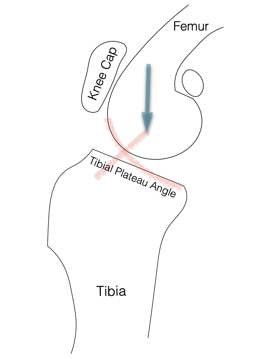
A torn CCL is a diagnosis made by a physical exam, not X-rays. However, X-rays are needed to demonstrate supportive evidence as well as evaluate joint swelling, the severity of osteoarthritis, joint angle (tibial plateau angle), and to rule out other causes of lameness. The three main physical exam findings most consistently identified in CCL rupture patients are joint swelling, instability (cranial drawer/cranial tibial thrust), and pain with hyperextension of the knee.

One of the largest differences between the human knee and the canine knee is the Tibial Plateau Angle (TPA). In humans, the TPA is 0-10 degrees, whereas in the dog the average TPA is about 25 degrees. During normal weight-bearing, the down forces through the knee are transmitted about perpendicular to the ground. (arrow).
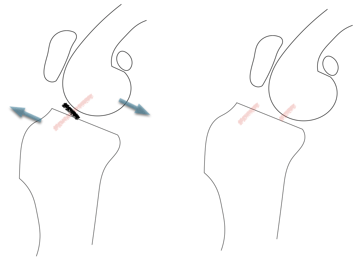
However, since the TPA is sloped down the round portion of the femur is encouraged to slide down the tibial plateau (cranial tibial thrust) This motion is prevented with a normal intact CCL. With a torn CCL, this instability (cranial tibial thrust) is not prevented allowing the round portion of the femur to slide down the slope.
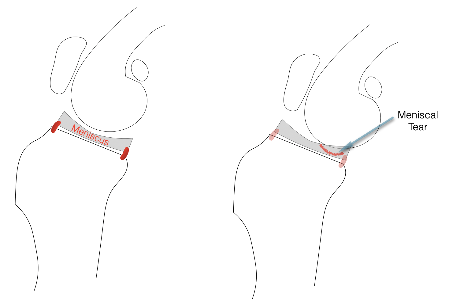
Instability in the knee from a torn CCL predisposes injury to a fibrocartilage structure between the femur and tibia called the medial meniscus. The meniscus functions to pick up the slack that is created from a round structure (femur) sitting on a flat surface (tibia). It helps to distribute forces over the joint surface more evenly. The reason the meniscus is at risk of damage is due to the fact that it is trapped on the top of the tibia by two ligaments. When instability from a torn CCL is present and the femur is allowed to move backward, a large amount of pressure is placed on the back portion of the medial meniscus which can result in a torn meniscus. Damage to the meniscus is evaluated and identified at the time of surgery. If there is a tear of the meniscus, the torn portion is removed. If the meniscus appears normal some surgeons will opt to release the meniscus while others choose to leave it intact. Even after the meniscus is evaluated and addressed at the time of surgery, there is a risk of future injury to the meniscus that could require a second surgery. This risk is small but will occur in about 5% of patients.
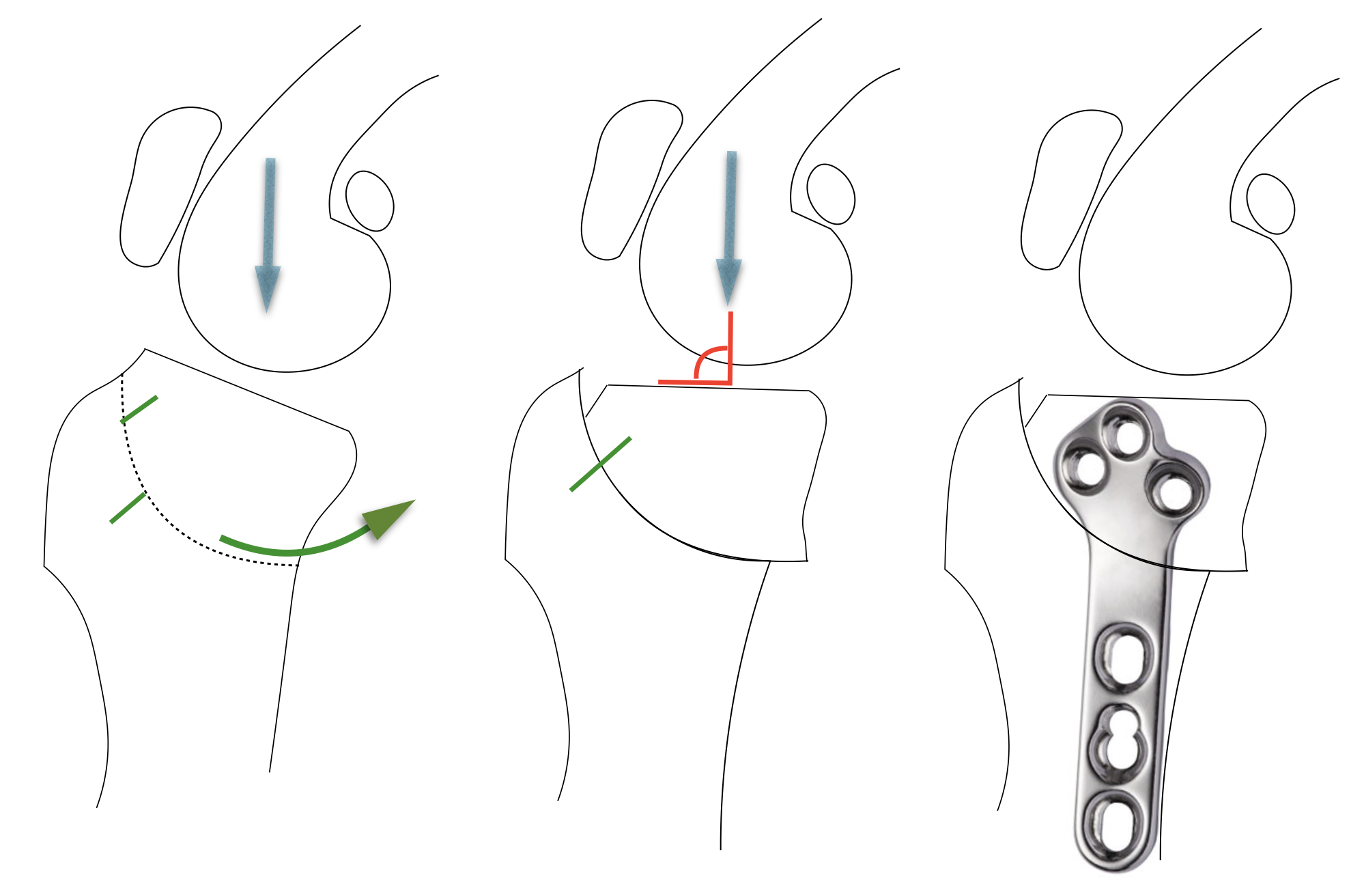
Once the joint has been “cleaned up” (torn CCL and meniscus addressed) we then move on to stabilizing the joint. There are several techniques available to repair the CCL. Two of the more common techniques used are the TPLO and MRIT (or Lateral Suture). The MRIT is a technique that utilizes a heavy non-absorbable suture and efficiently oriented scar tissue to create stability, whereas the TPLO requires a bone cut to stabilize the knee by altering the geometry in the knee. There are several factors that are considered when determining the best fit between the technique and the patient, including: size, age, activity level, anatomy, etc. The goal of the TPLO is to create weight-bearing stability by leveling the top of the tibia. A curved bone cut is made in the upper portion of the tibia, the tibial plateau is rotated along the curve (a calculated distance based on the slope of the tibial plateau) and stabilized in place with a bone plate and screws. By doing this the down forces of the leg are perpendicular to the tibial plateau which stabilizes the knee during weight-bearing.
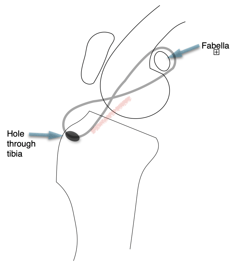
The MRIT (also called “lateral suture”, “extracapsular technique”, “fabella suture”) is a technique that depends upon the use of a heavy suture and development of scar tissue for stabilization of the knee. A non-absorbable suture is placed around the fabella and passed through a hole that has been drilled through the front of the tibia. The suture provides initial holding strength but can loosen over time. However, the suture acts as a scaffold for the body to create scar tissue. The scar tissue develops in an efficient orientation to resist instability and, therefore, stabilizes the knee.

Recovery

Recommendations for recovery from knee surgery can vary widely. Some encourage aggressive physical therapy starting the first week out of surgery and others focus on strict exercise restriction. Postoperative recommendations need to be tailored to each patient. In general, we recommend encouraging short stents of low impact weight-bearing on a leash for the first 6 weeks followed by increased duration and intensity of activity (on a leash) for the following 6 weeks. A radiograph is taken 10-12 weeks postoperatively to document bone healing for the TPLO. Once we have established a healed or progressively healing bone, higher impact activity can begin. It is not uncommon that intermittent lameness can be seen after trying new and more vigorous activities. Most dogs will work these “kinks” out during the fourth month postoperatively when higher impact activities begin.

Info on CCL Tears